



Services & Departments

Otorhinolaryngology (ENT)

Email: info@medihospital.com.cy

Telephone:

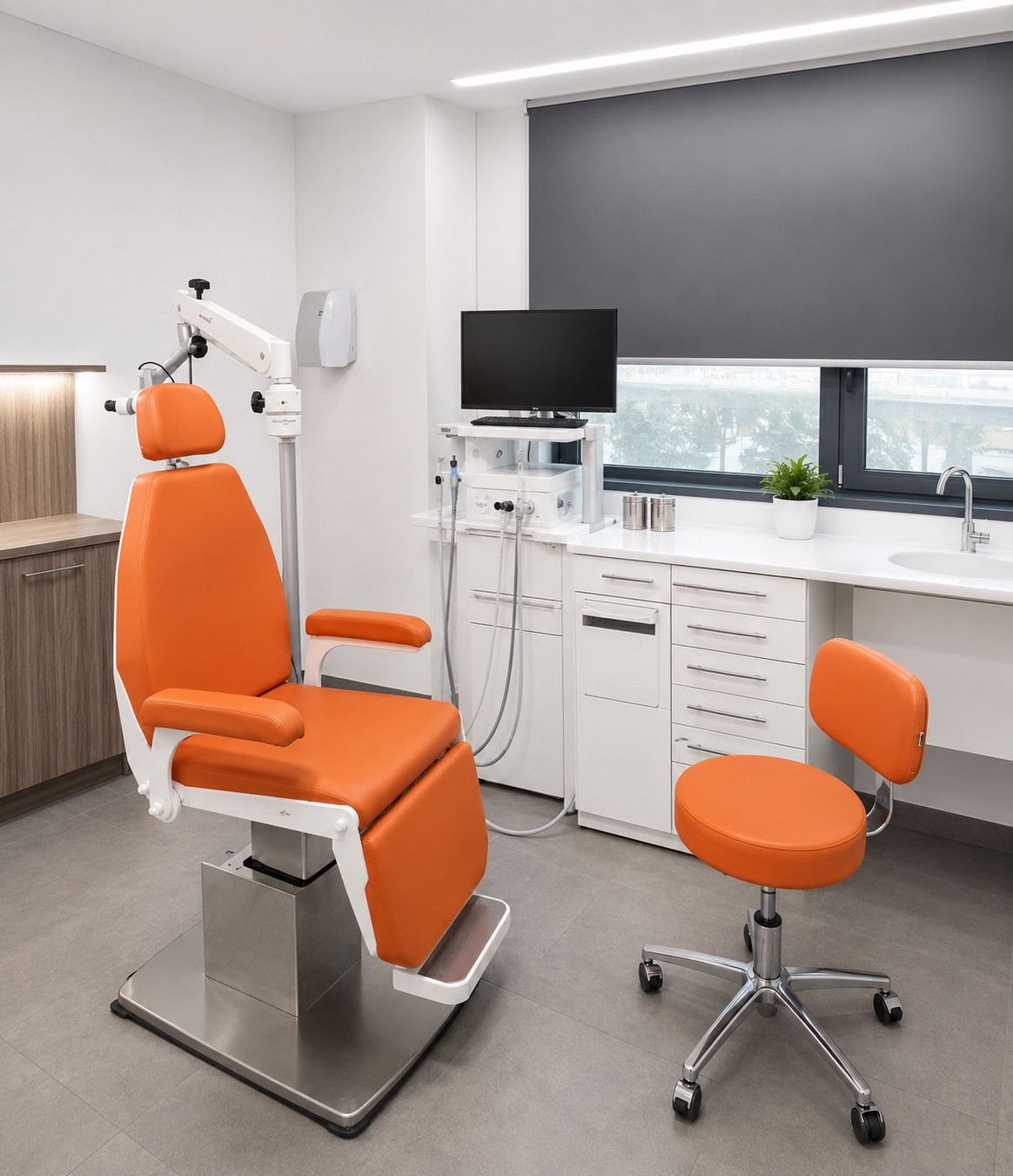
The Ear, Nose and Throat (ENT) Department at Mediterranean Hospital of Cyprus is a modern, specialised unit dedicated to the diagnosis, treatment and surgical management of head and neck conditions, providing comprehensive, high-quality services for both adults and children.

The Department covers the full spectrum of modern ENT care, including the management of:

  • Ear, nose and throat conditions
  • Hearing and balance disorders
  • Sinusitis and paranasal sinus conditions
  • Allergic and inflammatory conditions of the upper respiratory tract
  • Voice and laryngeal disorders
  • Sleep apnoea and snoring
  • Salivary gland disorders
  • Head and neck trauma
  • Head and neck tumours
  • Complex cases requiring specialised surgery and multidisciplinary management

The ENT Department operates dedicated outpatient clinics, providing:

  • Specialist ENT assessment
  • Endoscopic examination
  • Diagnostic evaluation
  • Preventive screening
  • Individualised treatment and follow-up

The Department also performs scheduled surgical procedures, using modern and minimally invasive techniques in:

  • Endoscopic nasal and sinus surgery
  • Ear surgery
  • Laryngeal and vocal cord surgery
  • Tonsil and adenoid surgery
  • Neck surgery
  • Head and neck surgery

The Department’s physicians actively participate in multidisciplinary meetings and clinical boards within the hospital for cases requiring a multidisciplinary approach, particularly in the areas of:

  • Head and neck oncology
  • Trauma
  • Neurosurgery
  • Paediatric cases
  • Complex surgical conditions

The ENT Department actively participates in the hospital’s on-call service, providing specialist ENT assessment and intervention for both inpatients and emergency cases.

A specialist ENT surgeon is on call 24 hours a day, 365 days a year, supporting the Accident and Emergency Department (A&E) and ensuring the prompt management of ENT emergencies.

The Department works in close collaboration with:

  • The Maxillofacial Surgery Department
  • The Neurosurgery Department
  • The Paediatrics Department
  • The Oncology Department
  • The Diagnostic Radiology Department
  • All other specialised surgical and clinical services within the hospital

This multidisciplinary approach ensures comprehensive and coordinated care tailored to the needs of each patient.
Το Τμήμα Ωτορινολαρυγγολογίας του Mediterranean Hospital of Cyprus διαθέτει σύγχρονες υποδομές και προηγμένο ιατροτεχνολογικό εξοπλισμό, επιτρέποντας την ακριβή διάγνωση και την ασφαλή διενέργεια εξειδικευμένων ΩΡΛ επεμβάσεων.

Το τμήμα διαθέτει:

  • σύγχρονες Χειρουργικές Αίθουσες
  • ενδοσκοπικά συστήματα υψηλής ευκρίνειας
  • συστήματα ενδοσκοπικής χειρουργικής ρινός και παραρρινίων
  • χειρουργικά μικροσκόπια
  • εξοπλισμό μικροχειρουργικής αυτιού
  • συστήματα διεγχειρητικής παρακολούθησης
  • προηγμένα συστήματα απεικόνισης
  • υπερηχογραφικά συστήματα
  • σύγχρονα αναισθησιολογικά και monitoring systems
  • καθώς και πλήρη διασύνδεση με το Ακτινοδιαγνωστικό Τμήμα και τις μονάδες εντατικής φροντίδας

Η λειτουργία του τμήματος υποστηρίζεται επίσης από:

  • Αξονικούς Τομογράφους 160 τομών
  • Μαγνητικούς Τομογράφους 1.5 Tesla
  • PACS & EMR συστήματα
  • προηγμένα ακτινοδιαγνωστικά συστήματα
  • καθώς και πλήρεις υποδομές χειρουργικής και μετεγχειρητικής υποστήριξης

Η προηγμένη τεχνολογική υποδομή επιτρέπει:

  • ακριβέστερη διάγνωση
  • αυξημένη χειρουργική ακρίβεια
  • εφαρμογή ελάχιστα επεμβατικών τεχνικών
  • μείωση του χειρουργικού τραύματος
  • ταχύτερη ανάρρωση
  • καθώς και βελτιωμένα λειτουργικά και αισθητικά αποτελέσματα για τους ασθενείς

The ENT Department is staffed by specialist ENT surgeons with extensive experience across the full spectrum of modern otolaryngology and head and neck surgery.

The medical team works closely with:

  • Anaesthesiologists
  • Neurosurgeons
  • Maxillofacial Surgeons
  • Oncologists
  • Paediatricians
  • Radiologists
  • All other specialised services within the hospital

This close collaboration ensures a comprehensive, multidisciplinary approach to the care of every patient.

The specialised nursing team has extensive experience in:

  • ENT surgical procedures
  • Postoperative care
  • Management of emergency cases
  • Care and support of patients with complex head and neck conditions

Continuous professional development, adherence to international clinical guidelines and the use of advanced technologies reflect Mediterranean Hospital of Cyprus’ commitment to providing high-quality ENT services, with a strong focus on safety, quality and compassionate patient care.

Services

Parathyroidectomy

Parathyroidectomy is a surgical procedure involving the removal of one or more parathyroid glands. The procedure is indicated for patients with a parathyroid adenoma, hyperparathyroidism or parathyroid cancer.

When a single parathyroid gland is affected, most commonly in patients with primary or tertiary hyperparathyroidism, only the affected gland (adenoma) is removed. The removal of a single abnormal parathyroid gland (parathyroid adenoma) accounts for the vast majority of parathyroidectomy procedures.

Parathyroidectomy is performed under general anaesthesia through a small transverse incision in the lower part of the neck. Ideally, the surgeon places the incision along a pre-existing natural skin crease. In exceptional cases, most commonly when bilateral neck exploration is indicated, the incision may need to be extended by approximately one to two centimetres.

In cases of a solitary parathyroid adenoma, the endocrine surgeon initially approaches the side on which the abnormal gland has been identified. The surgeon then locates the parathyroid adenoma and carefully removes it.

In patients with diffuse hyperplasia involving all parathyroid glands, the surgeon performs bilateral neck exploration to identify all the parathyroid glands. This is followed by either a subtotal parathyroidectomy or a total parathyroidectomy with autotransplantation of a portion of parathyroid tissue.

At the end of the procedure, the skin incision is closed with an intradermal suture, helping to achieve an excellent cosmetic result.

Thyroidectomy

Thyroid surgery may be necessary in the following cases:

  • When one or more thyroid nodules are present and there is significant suspicion of thyroid cancer.
  • In cases of diffuse enlargement of the thyroid gland (goitre), when it:
    • Compresses the trachea and oesophagus, causing difficulty in breathing and swallowing.
    • Causes cosmetic concerns due to its size.
    • Is suspected of containing cancerous tissue.
  • In cases of excessive production of thyroid hormones (hyperthyroidism) that cannot be adequately controlled with medication.

The procedure is performed under general anaesthesia through a small incision at the front of the neck, just above the thyroid gland. Whenever possible, the incision is placed within a natural skin crease of the neck. Careful closure using plastic surgery techniques at the end of the procedure helps achieve an excellent cosmetic result.

In a total thyroidectomy, the entire thyroid gland is removed, while in a partial thyroidectomy, one lobe of the thyroid, either the right or the left, is usually removed.

Of particular importance during surgery is the identification and preservation of the anatomical and functional integrity of the laryngeal nerves, including the recurrent and superior laryngeal nerves, which are responsible for vocal cord function.

The use of specialised intraoperative nerve monitoring significantly reduces the risk of injury to these nerves.

It is also essential to identify and preserve the parathyroid glands. These are four small glands located behind the thyroid gland that play a vital role in regulating calcium and phosphorus levels in the body.

The usual hospital stay is 1–2 days.

A small drainage tube placed at the incision site is usually removed the day after surgery. Blood calcium levels are checked on the evening of the operation and again the following day. The sutures used are generally absorbable and therefore do not need to be removed. Patients can usually return to work within two weeks.

Following a total thyroidectomy, lifelong thyroid hormone replacement therapy in tablet form is required to replace the hormones normally produced by the thyroid gland.

Minor Salivary Gland Biopsy

In certain rheumatological and autoimmune conditions, such as Sjögren’s syndrome, early and accurate diagnosis is particularly important for appropriate patient monitoring and treatment. One of the investigations that can play an important role in the diagnostic process is a minor salivary gland biopsy.

The minor salivary glands are distributed throughout the oral cavity and are particularly abundant in the lower lip. By obtaining a small tissue sample, the physician can examine it microscopically for inflammatory changes associated with autoimmune conditions.

The procedure is usually performed under local anaesthesia and is quick, safe and minimally invasive. A small incision is made on the inner surface of the lower lip, through which a few minor salivary glands are removed for histological examination. The area is closed with fine absorbable sutures, and the patient can usually return to normal daily activities within a short period of time.

The procedure is generally performed as a day-case procedure and does not require an overnight hospital stay, allowing the patient to be discharged on the same day.

At Mediterranean Hospital, the procedure is performed by a specialised medical team experienced in the diagnosis and treatment of conditions affecting the oral cavity and salivary glands, with particular emphasis on patient safety, comfort and individualised care.

Submandibular Gland Surgery

The submandibular glands are among the major salivary glands. They are located beneath the jaw, with one gland on each side.

Their function is to produce saliva, which is carried through a small tube (duct) into the oral cavity.

The most common problem affecting the submandibular salivary gland is obstruction of its duct. This may be caused by narrowing of the duct or by the formation of a small stone (salivary stone), as the saliva produced by the submandibular gland is relatively thick.

This condition typically presents as sudden, painful swelling of the gland, usually after eating. In some cases, the swelling resolves on its own. In others, however, it may lead to persistent inflammation requiring treatment with antibiotics and anti-inflammatory medication.

As with the parotid gland, tumours may also develop within the submandibular salivary glands. A significant proportion of these tumours may be malignant (cancerous).

Surgery is performed under general anaesthesia through a small incision, which is placed within a natural skin crease of the neck whenever possible. Careful closure using plastic surgery techniques at the end of the procedure helps achieve an excellent cosmetic result. The procedure takes approximately one hour.

The usual hospital stay is 1–2 days. A small drainage tube placed at the incision site is generally removed the day after surgery, while the sutures are removed after approximately 12–15 days. Patients can usually return to work within two weeks.

Parotidectomy

The parotid glands are the largest salivary glands in the human body. There are two parotid glands, one on each side of the face, located in front of and below each ear. Their function is to produce saliva, which is carried through a small tube (duct) into the oral cavity. In some cases, cysts or tumours may develop within the parotid gland. Approximately 80% of parotid tumours are benign, while malignant tumours (cancer) in this area are less common.

Modern parotid surgery involves removing not only the tumour but also an appropriate margin of surrounding healthy tissue in order to reduce the risk of recurrence. The procedure is performed under general anaesthesia, using an incision similar to that used in facelift surgery. Careful closure using plastic surgery techniques at the end of the procedure helps achieve an excellent cosmetic result.

Of particular importance during surgery is the identification and preservation of the anatomical and functional integrity of the facial nerve. This nerve controls the muscles responsible for facial movement and passes through the parotid gland. The use of specialised intraoperative nerve monitoring to identify and monitor the main trunk and branches of the facial nerve significantly reduces the risk of nerve injury.

The usual hospital stay is 1–2 days. A pressure dressing is applied to the area and is kept in place for several days, with dressing changes and wound care carried out during follow-up visits. A small drainage tube placed at the incision site is usually removed after 2–3 days, while the sutures are removed after approximately 12–15 days. Patients can usually return to work within two weeks.

Vocal Cord Surgery

Under normal conditions, the vocal cords are smooth and regular and move symmetrically, producing a clear, natural voice that reflects their healthy function. In some cases, however, this normal function may be disrupted, causing the voice to become hoarse, weak or easily fatigued during speaking. Some patients may even report that their voice fades or is completely lost after only a short period of speaking.

Depending on the underlying cause of the voice disorder, treatment may be either conservative or surgical. Conservative treatment may include medication, specialised advice on vocal hygiene, speech and voice therapy, and voice training. When surgical intervention is required, modern vocal cord microsurgery allows lesions to be removed with exceptional precision while preserving the delicate anatomical structures of the larynx.

Conditions that can be treated with vocal cord microsurgery include:

  • Vocal cord polyps
  • Vocal cord nodules
  • Vocal cord cysts
  • Polypoid degeneration (Reinke’s oedema)
  • Granulomas
  • Papillomas
  • Haemangiomas
  • Leukoplakia, a potentially precancerous condition
  • Early-stage, localised carcinomas

The procedure is performed under general anaesthesia using a surgical laryngoscope, without the need for any external incision. To achieve the highest possible degree of precision, a specialised surgical microscope or high-definition endoscope is used.

The lesion is removed using specialised laryngeal microsurgical instruments or a high-precision surgical laser. This advanced technology provides excellent surgical precision and effectiveness, while ensuring maximum preservation of the delicate anatomical structures of the vocal cords and larynx, helping to maintain and restore voice quality.

Φαρυγγοπλαστική

Η υπερτροφία ή χαλάρωση των μαλακών ιστών του φάρυγγα, όπως είναι οι αμυγδαλές, η σταφυλή και η υπερώα (ουρανίσκος), μπορεί να προκαλούν ή να επιδεινώνουν το ροχαλητό ή την υπνική άπνοια.

Η φαρυγγοπλαστική είναι μία επέμβαση, κατά την οποία αφαιρούνται οι αμυγδαλές ή μειώνεται ο όγκος τους, μειώνεται το μήκος της σταφυλής, και με ειδικές τομές στο πλάι της σταφυλής μειώνεται το δονούμενο τμήμα της υπερώας. Η επέμβαση γίνεται με γενική αναισθησία και με τη βοήθεια των ραδιοσυχνοτήτων(RF) ή του Laser.

Διαρκεί περίπου 30 λεπτά και ο ασθενής μπορεί να επιστρέψει στο σπίτι του την ίδια ημέρα εφόσον δεν αφαιρούνται οι αμυγδαλές.
Βελτίωσης της ρινικής αναπνοής: ευθειασμός ρινικού διαφράγματος, κογχοτομές, καυτηριασμός ρινικών κογχών, ενδοσκοπική χειρουργική ρινός παραρρινίων – αφαίρεση ρινικών πολυπόδων.
Φαρυγγοϋπερωιοσταφυλοπλαστική (UPPP) είναι η βασική επέμβαση για το ροχαλητό και το απνοϊκό σύνδρομο. Αφαιρούνται πληθωρικοί ιστοί του φάρυγγα (ευμεγέθης σταφυλή, μεγάλες παρίσθμιες καμάρες, τοιχώματα με πτυχώσεις).

Πιο σύγχρονη παραλλαγή φαρυγγοπλαστικής UPPP με χρήση laser και κυρίως ραδιοσυχνοτήτων (RF). Οι ραδιοσυχνότητες μπορεί να χρησιμοποιηθούν και προκαλώντας ουλοποίηση της μαλθακής υπερώας αυξάνοντας την ακαμψία της και μειώνοντας έτσι τις δονήσεις και το ροχαλητό.

Αδενοτομή (Κρεατάκια)

Οι αδενοειδείς εκβλαστήσεις, τα γνωστά σε όλους κρεατάκια βρίσκονται στο ρινοφάρυγγα ακριβώς πίσω από τη μύτη. Πιστεύεται ότι μέχρι την ηλικία των 5-6 ετών παίζουν κάποιο ρόλο στην άμυνα στην οργανισμού ενώ έχουν την τάση να συρρικνώνονται και προοδευτικά να εξαφανίζονται μέχρι την ενήλικη ζωή. Σε κάποιες όμως περιπτώσεις το μέγεθός τους είναι μεγαλύτερο από το φυσιολογικό με αποτέλεσμα ο ρινοφάρυγγας να ‘κατακλύζεται’ από τα κρεατάκια γεγονός που έχει μια σειρά από αρνητικές επιπτώσεις:
Έτσι το παιδί με υπερτροφικές αδενοειδείς εκβλαστήσεις δεν μπορεί να αναπνεύσει από τη μύτη και καταφεύγει στην αναπνοή από το στόμα, κάτι που σε βάθος χρόνου μπορεί να οδηγήσει σε παραμόρφωση των οστών της γνάθου και του κρανίου, το λεγόμενο ‘αδενοειδές προσωπείο’.

Κατά τη διάρκεια του ύπνου το παιδί ροχαλίζει και κάνει άπνοιες δηλ. σταματάει η αναπνοή του για μερικά δευτερόλεπτα. Αυτή η κακή ποιότητα του ύπνου προκαλεί κούραση και υπνηλία ή υπερδιέγερση κατά τη διάρκεια της ημέρας, διάσπαση προσοχής και κακές επιδόσεις στο σχολείο, ακόμη και μαθησιακές δυσκολίες. Η σοβαρή άπνοια μάλιστα αν δεν διαγνωστεί και αντιμετωπιστεί έγκαιρα είναι δυνατόν σε βάθος χρόνου να οδηγήσει σε σοβαρά καρδιοαναπνευστικά προβλήματα.

Το παιδί μπορεί να έχει βαρηκοΐα και συχνές ωτίτιδες εξαιτίας του υγρού που μαζεύεται πίσω από το τύμπανο του αυτιού.Τέλος, σε κάποιες περιπτώσεις, τα κρεατάκια αποτελούν μία ‘δεξαμενή’ (‘reservoir’) μικροβίων, γεγονός που προδιαθέτει σε συχνές, υποτροπιάζουσες λοιμώξεις με εκροή πυώδους βλέννας από τη μύτη και βήχα, που ανθίστανται στην αντιβιοτική αγωγή.

Σε όλες τις παραπάνω περιπτώσεις η χειρουργική επέμβαση της αδενοτομής, η αφαίρεση δηλαδή των αδενοειδών εκβλαστήσεων, λύνει οριστικά το πρόβλημα και εξασφαλίζει στο παιδί τόσο μια ομαλή καθημερινότητα όσο και μια φυσιολογική ανάπτυξη.

Αμυγδαλεκτομή

Υποψήφιοι για να υποβληθούν σε αμυγδαλεκτομή είναι παιδιά ή ενήλικες με:

  • Συχνές υποτροπιάζουσες αμυγδαλίτιδες.
  • Ιστορικό περιαμυγδαλικού αποστήματος.
  • Προσβολή των νεφρών και της καρδιάς ως αποτέλεσμα της διασποράς των μικροβίων στο αίμα εξαιτίας της αμυγδαλίτιδας.
  • Υπερτροφικές αμυγδαλές που προκαλούν ροχαλητό και άπνοια κατά τη διάρκεια του ύπνου.
  • Δυσοσμία του στόματος.
  • Υποψία κακοήθειας.

Η επέμβαση γίνεται υπό γενική αναισθησία είτε με τη χρήση κλασικών εργαλείων και διαθερμίας είτε με τη χρήση άλλων αναίμακτων τεχνικών όπως coblation ή ligasure. Πριν την επέμβαση είναι απαραίτητος ο προεγχειρητικός έλεγχος (εξετάσεις αίματος, εκτίμηση από καρδιολόγο και αναισθησιολόγο) ο οποίος στις περισσότερες περιπτώσεις γίνεται την ημέρα του χειρουργείου, λίγες ώρες πριν την έναρξη της επέμβασης.

Η διάρκειά της είναι συνήθως γύρω στα 30-60 λεπτά.

Τοποθέτηση Σωληνίσκων Αερισμού

Υπό φυσιολογικές συνθήκες, ο χώρος που βρίσκεται πίσω από το τύμπανο του αυτιού είναι γεμάτος με αέρα, γεγονός που εξασφαλίζει την απρόσκοπτη μετάδοση του ηχητικού κύματος και κατ’ επέκταση τη φυσιολογική ακοή. Σε κάποιες όμως περιπτώσεις, ο χώρος αυτός γεμίζει με υγρό, άλλοτε λεπτόρευστο και άλλοτε παχύρευστο. Η κατάσταση αυτή επιστημονικά ονομάζεται εκκριτική ωτίτιδα και στη συντριπτική πλειοψηφία των περιστατικών αφορά παιδιά.

Ο μικρός ασθενής δεν αισθάνεται κάποιο πόνο όμως η ακοή του επηρεάζεται σημαντικά καθώς αυτή μπορεί να μειωθεί έως και κατά 35 dB. Το ζήτημα είναι ότι τα περισσότερα παιδιά δεν μπορούν να αντιληφθούν και να επικοινωνήσουν το πρόβλημά τους και έτσι έγκειται στους γονείς να καταλάβουν ότι το παιδί τους πάσχει από βαρηκοΐα. Σημάδια που μπορούν να αποκαλύψουν κάτι τέτοιο είναι ένα παιδί που δεν ακούει όταν του μιλάμε και δείχνει αφηρημένο ή απορροφημένο σε αυτό που κάνει καθώς επίσης και το υπερβολικό δυνάμωμα του ήχου της τηλεόρασης. Η απόδοση στο σχολείο πολλές φορές δεν είναι καλή ενώ στις περιπτώσεις που το υγρό είναι παρόν από τη βρεφική ηλικία καθυστερεί η ανάπτυξη του λόγου και η άρθρωση των λέξεων επηρεάζεται σημαντικά.

Η αρχική αντιμετώπιση της εκκριτικής ωτίτιδας είναι συνήθως συντηρητική με ειδικές ασκήσεις που δίνονται στο παιδί, ρινοπλύσεις και παρακολούθηση. Αν αυτή δεν αποδώσει, τότε κάνουμε παρακέντηση του τυμπάνου και αφαιρούμε το υγρό ενώ στις περισσότερες περιπτώσεις, προκειμένου να αποτρέψουμε την υποτροπή του υγρού τοποθετούμε στο τύμπανο σωληνίσκο αερισμού. Η επέμβαση γίνεται με μέθη ή με γενική αναισθησία και συνήθως διαρκεί 5 με 10 λεπτά. Η αποκατάσταση της ακοής στο φυσιολογικό είναι άμεση. Αν συνυπάρχει και υπερτροφία των αδενοειδών εκβλαστήσεων (κρεατάκια), αυτά αφαιρούνται στην ίδια επέμβαση καθώς έχει βρεθεί ότι συμβάλλουν στη συλλογή υγρού πίσω από το τύμπανο.

Ωτοπλαστική

Ωτοπλαστική ονομάζεται η χειρουργική επέμβαση που γίνεται στα αυτιά, προκειμένου να διορθώσουμε το σχήμα, τη θέση ή το μέγεθός τους. Με αυτή την επέμβαση πολύ συχνά αντιμετωπίζονται ταυτόχρονα τόσο λειτουργικά, όσο και αισθητικά προβλήματα που προκύπτουν από την μορφή των αυτιών.

Η επέμβαση πραγματοποιείται στο εξωτερικό τμήμα τους, το λεγόμενο ωτικό πτερύγιο.
Το πτερύγιο αποτελείται από πτυχές χόνδρινου ιστού καλυμμένες από δέρμα. Η ανάπτυξή του αρχίζει πριν τη γέννηση και συνεχίζεται στα πρώτα χρόνια της παιδικής ηλικίας.

Αν η ανάπτυξη αυτή δε γίνει όπως πρέπει, τότε μπορούμε να επιτύχουμε τη αναδιαμόρφωση του ωτικού χόνδρου με τη χειρουργική επέμβαση της ωτοπλαστικής.

Τα πεταχτά αυτιά ή σύμφωνα με την επιστημονική ορολογία αφεστώτα ώτα αποτελούν μία από τις συχνότερες γενετικές δυσμορφίες του προσώπου (5% περίπου του γενικού πληθυσμού). Τη συναντούμε τόσο σε αγόρια όσο και σε κορίτσια ενώ το πρόβλημα που προκαλεί είναι καθαρά αισθητικό.

Έτσι δεν είναι λίγες οι περιπτώσεις που ένα παιδί με πεταχτά αυτιά αισθάνεται μειονεκτικά όσον αφορά την εμφάνισή του, κάτι που αν δεν διορθωθεί έγκαιρα θα επηρεάσει σημαντικά την αυτοπεποίθησή του και πιθανότατα την καθημερινότητά του στο μέλλον. Αρκετές φορές μάλιστα τα παιδιά αυτά γίνονται αντικείμενο άκομψων πειραγμάτων στο σχολείο και στις παρέες κάτι που μπορεί να επηρεάσει πολύ σημαντικά την ψυχολογία τους.

Department Doctors

Director

Otorhinolaryngology

Otorhinolaryngology

Otorhinolaryngology

Otorhinolaryngology